[最も好ましい] e coli under microscope 10x 148061-What does e coli look like under a microscope
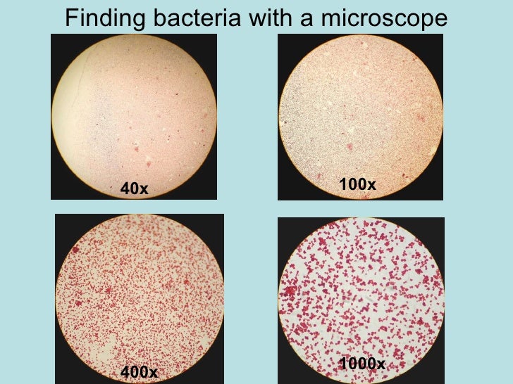
7 NOW, switching to the 40X objective with the 10X eyepiece gives 400X magnification The field of view is This is illustrated by the 400X circle on the 100X view This circle is ¼ the diameter of the 100X view 8 Switch to the 400X view and 400X ruler (a millimeter ruler as seen under 400 power magnification)Which bacteria look similar to E coli under 100X optical microscope?First, by using Escherichia coli (Ecoli), we start at 4x objective, followed by low power 10x objective viewing and the specimen looked clearer at 40x magnificationThe shaped of Ecoli is rodshaped and it is a gramnegative bacteriaThe gram stain of some specimen either gramnegative or grampositive can be resulted by looked at their color whether the bacteria are pink or red for gram
Q Tbn And9gcqkye60ou Johpr02n Mbv1fferrjpdh Lnct7ymdf5qhyia1ld Usqp Cau
What does e coli look like under a microscope
What does e coli look like under a microscope-Bacteria E coli Must observe under 400x Very small & motile Looks like specks of sand Hard to discern shape Smaller than yeast & protozoaMost educationalquality microscopes have a 10x (10power magnification) eyepiece and three objectives of 4x, 10x & 40x to provide magnification levels of 40x, 100x and 400x (ii) Another thing to consider is that microscope resolution which is the ability to see close but separate points as distinct comes from the objective lenses, not from


Flower Like Patterns In Multi Species Bacterial Colonies Elife
2 Negative endospore stain showing only vegetative cells @1000xTM;4 Can a light microscope see bacteria?To observe E coli with any detail, you will need to use the 100X lens, which is also known as an oil immersion lens This is the longest, most powerful and most expensive lens on the microscope, requiring extra care when using it As the name implies, the 100X lens is immersed in a drop of oil on the slide
Most of the bacteria range from 022 µm in diameter The length can range from 110 µm for filamentous or rodshaped bacteria The most wellknown bacteria E coli, their average size is ~15 µm in diameter and 26 µm in length In this figure The size comparison between our hair (~ 60 µm) and E coli (~1 µm) Notice how small theLike E coli live in the intestines of animals where they help in producing the vitamin k2 in addition to preventing pathogenic bacteria from thriving in the large intestine See Also EColi Under the MicroscopeE coli under the microscope Matthew Tessnear Tuesday Oct 16, 12 at 11 AM Oct 16, 12 at 525 PM Health officials say they expect a slow down soon in the number of reported new E coli cases in an outbreak linked to the Cleveland County Fair
Lens at the top of microscope 🔬 that users lookEntamoeba histolytica is an anaerobic parasitic amoebozoan, part of the genus Entamoeba Predominantly infecting humans and other primates causing amoebiasisEscherichia coli (/ ˌ ɛ ʃ ə ˈ r ɪ k i ə ˈ k oʊ l aɪ /), also known as E coli (/ ˌ iː ˈ k oʊ l aɪ /), is a Gramnegative, facultative anaerobic, rodshaped, coliform bacterium of the genus Escherichia that is commonly found in the lower intestine of warmblooded organisms (endotherms) Most E coli strains are harmless, but some serotypes (EPEC, ETEC etc) can cause serious food


Interdependency Of Formation And Localisation Of The Min Complex Controls Symmetric Plastid Division Journal Of Cell Science


Characterization Of Biofilm Forming Capacity And Resistance To Sanitizers Of A Range Of E Coli O26 Pathotypes From Clinical Cases And Cattle In Australia Bmc Microbiology Full Text
Examples of Bacteria Under the Microscope Escherichia coli Escherichia coli (Ecoli) is a common gramnegative bacterial species that is often one of the first ones to be observed by students Most strains of Ecoli are harmless to humans, but some are pathogens and are responsible for gastrointestinal infections They are a bacillus shapedA rectangular piece of glass where a sample is mounted for viewing under the microscope What is the eye piece?Find yeast microscope stock images in HD and millions of other royaltyfree stock photos, illustrations and vectors in the collection Thousands of new, highquality pictures added every day


Q Tbn And9gcstf8rlsv6xf Tlmjmgapkp5vctdrjr4c7vz0kjmptie3nty0xo Usqp Cau


Microscopy For The Winery Viticulture And Enology
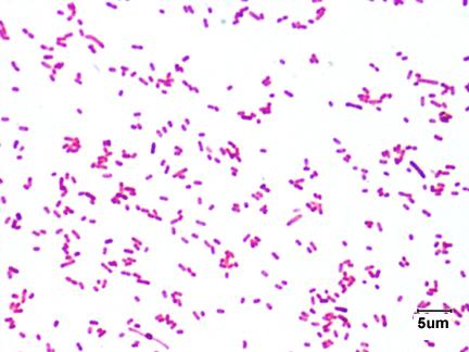
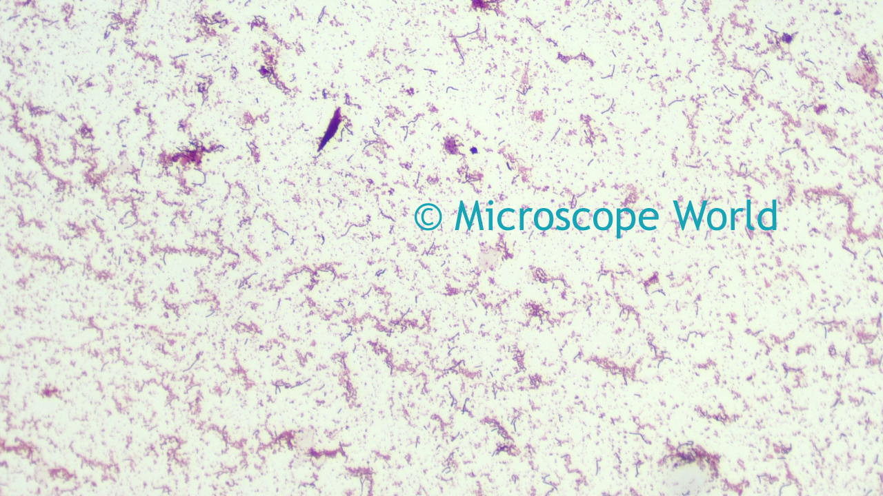
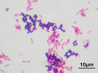
E coli are particularly useful to molecular biologists because they rapidly grow to very high densities in laboratory culture media, reaching densities of 1 10 billion cells per mL Although E coli are times smaller than yeast cells, their sheer numbers and their distinct motion renders them visible with the light microscope3 Malachite green primary staining step of endopore stain with slide being heated over water bath;Mixed Gram & Gram Bacteria Under Microscope (Click on image to enlarge) Gram stain mixed sample od Staphylococcus (Gram , purple cocci) & E coli , (Gram pink rods) @ 1000xTM


Gram Negative Synergy And Mechanism Of Action Of Alkynyl Bisbenzimidazoles Scientific Reports


Laboratory Test 1 Flashcards Chegg Com
Pick up a "tiny" amount of an Escherichia coli colony Focus on the line with the 10X objective – refer to the microscope focusing procedure described in lab 1 Once you have focused on the specimen using the 10X objective, move the 40X objective lens into position Use the fine adjustment knob to bring the specimen into focusMany bacteria look like E coli when examined under the microscope (if not stained Enterobacteriaceae, Bacillus, cornyeformeEndospore stained slide, with


Biol 230 Lab Manual Lab 1


Google Answers Microscopes
Find the perfect E Coli Under A Microscope stock photos and editorial news pictures from Getty Images Select from premium E Coli Under A Microscope of the highest quality• A basic light microscope has 4 objective lenses – 4X, 10X, 40X, and 100X The higher the number, the higher the power of magnification Once you have focused theLike E coli live in the intestines of animals where they help in producing the vitamin k2 in addition to preventing pathogenic bacteria from thriving in the large intestine See Also EColi Under the Microscope


The Crohn S Disease Associated Escherichia Coli Strain Lf Relies On Sos And Stringent Responses To Survive Multiply And Tolerate Antibiotics Within Macrophages
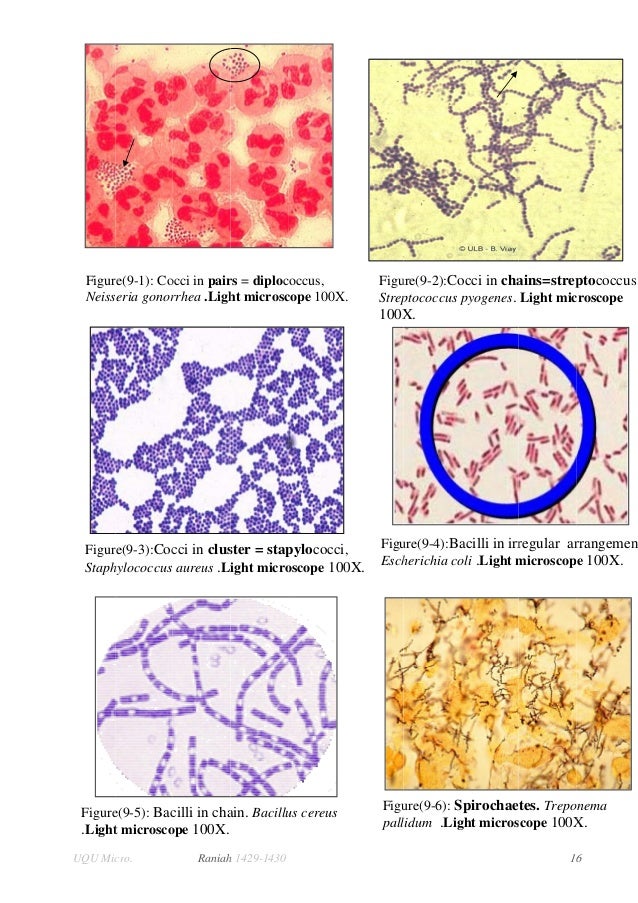


Lab 2 Lab 3
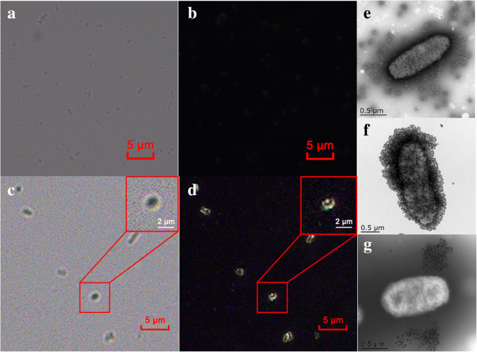
7 NOW, switching to the 40X objective with the 10X eyepiece gives 400X magnification The field of view is This is illustrated by the 400X circle on the 100X view This circle is ¼ the diameter of the 100X view 8 Switch to the 400X view and 400X ruler (a millimeter ruler as seen under 400 power magnification)Mixed Gram & Gram Bacteria Under Microscope (Click on image to enlarge) Gram stain mixed sample od Staphylococcus (Gram , purple cocci) & E coli , (Gram pink rods) @ 1000xTMEcoli is usually motile in liquid or semisolid environment with peritrichous flagella (about 6 per cell) and its surface is covered with fimbriae These structures (flagella and fimbriae) are too thin to be visualized by classical light microscopy or they don't have to be present at all under given cultivation conditions even at motile strains


Biol 230 Lab Manual Lab 1


Aph162 Report 1
Escherichia coli (E coli) bacteria normally live in the intestines of people and animals Most E coli are harmless and actually are an important part of a healthy human intestinal tract However, some E coli are pathogenic, meaning they can cause illness, either diarrhea or illness outside of the intestinal tract The types of E coli that can cause diarrhea can be transmitted throughEntamoeba coli E coli cysts in concentrated wet mounts Cysts of Entamoeba coli are usually spherical but may be elongated and measure 10–35 µm Mature cysts typically have 8 nuclei but may have as many as 16 or more Entamoeba coli is the only Entamoeba species found in humans that has more than four nuclei in the cyst stage The nuclei may be seen in unstained as well as stainedYes, most of the bacteria range from 022 µm in diameter The length can range from 110 µm for filamentous or rodshaped bacteria The most wellknown bacteria E coli, their average size is ~15 µm in diameter and 26 µm in length As we talked above, you can see some bacteria in my cheek cells


Gram Stain


Vade Mecum Microscope Practical Microscopy To Take With You Page 7
E coli under the microscope Escherichia coli (E coli) is a bacterium commonly found in various ecosystems like land and water Most of the strains of E coli are harmless, but some strains are known to cause diarrhea and even UTIs E coli is commonly studied as they are considered as a standard for the study of different bacteriaEscherichia coli bacteria on blood agar e coli under a microscope stock pictures, royaltyfree photos & images petri dishes with culture media for sarscov2 diagnostics, test coronavirus covid19, microbiological analysis e coli under a microscope stock pictures, royaltyfree photos & imagesWhat is the approximate size of Ecoli?


Q Tbn And9gcqkye60ou Johpr02n Mbv1fferrjpdh Lnct7ymdf5qhyia1ld Usqp Cau


What Does An E Coli Bacteria Look Like Under A Microscope Quora
Focusing With The 10X Objective Olympus CX31 Microscope 1 Rotate the yellowstriped 10X objective until it locks into place This will give a total magnification of 100X 2 Using the iris diaphragm lever under the stage (see Fig 6A), reduce the light by sliding the lever most of the way to the right 3Staphylococcus aureus and Escherichia coli under microscope, morphology and microscopic appearance Bacteria under Microscope Staphylococcus epidermidis Bacteria under Microscope Gramstain Grampositive Microscopic appearance Cocci in grapelike clusters, diplococci, cocci4 Observe Click on the cow and observe E coli under the highest magnification Notice the microscope magnification is larger for this organism, and notice the scale bar is smaller A What is the approximate size of E coli?


Direct E Coli Cell Count At Od600 Tip Biosystems


Biol 230 Lab Manual Lab 1
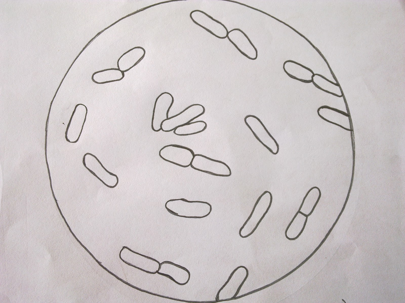
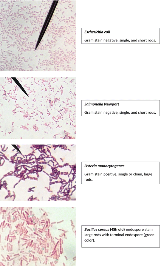
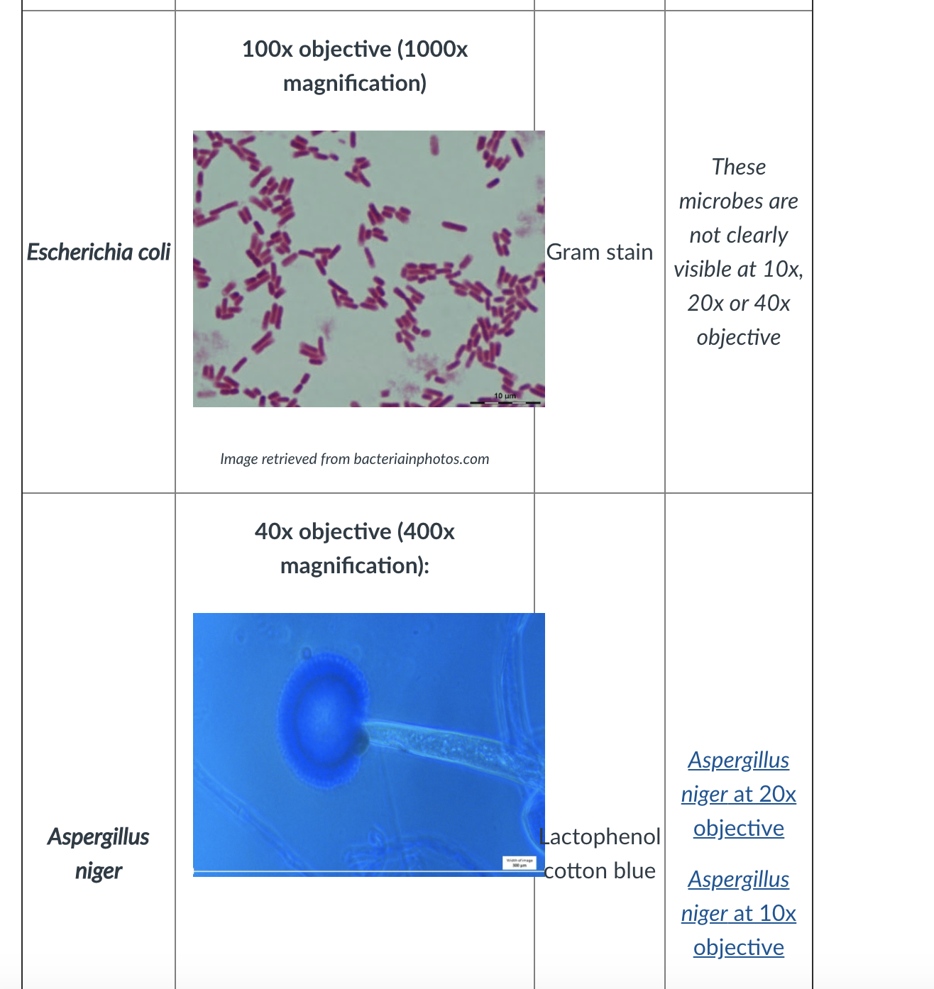
E coli bacteria looks like small rods under compound microscope at 1000 magnification power It is gram negative bacterium which appears pink when stained by a staining technique called as Gram staining which is a differential staining technique 1K viewsE coli under microscope By Barbara Duckworth Reading Time 3 minutes Published October 12, 00 Livestock The quest to unravel the mystery of E coli bacteria grows more perplexingEntamoeba coli E coli cysts in concentrated wet mounts Cysts of Entamoeba coli are usually spherical but may be elongated and measure 10–35 µm Mature cysts typically have 8 nuclei but may have as many as 16 or more Entamoeba coli is the only Entamoeba species found in humans that has more than four nuclei in the cyst stage The nuclei may be seen in unstained as well as stained
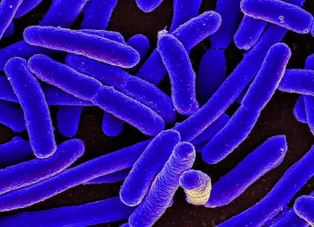


E Coli Under The Microscope Types Techniques Gram Stain Hanging Drop Method
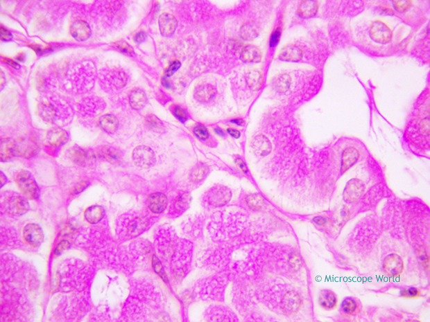


Why Would You Need Microscope Immersion Oil And How To Use It
Ecoli is usually motile in liquid or semisolid environment with peritrichous flagella (about 6 per cell) and its surface is covered with fimbriae These structures (flagella and fimbriae) are too thin to be visualized by classical light microscopy or they don't have to be present at all under given cultivation conditions even at motile strainsWhen viewed under the microscope, Gramnegative E Coli will appear pink in color The absence of this (of purple color) is indicative of Grampositive bacteria and the absence of Gramnegative E Coli Escherichia coli under 10х90х magnification using fuchsine as a dye by ElNokko (Own work) CC BYSA 40 (http//creativecommonsorg/licenses/bysa/40), via Wikimedia CommonsE Coli (Escherichia Coli) is a gramnegative, rodshaped bacterium Most E Coli strains are harmless, but some serotypes can cause food poisoning in their hosts The harmless strains are part of the normal flora of the gut Learn more about E Coli here Helicobacter Pylori The prepared microscope slide image of Helicobacter Pylori at left was captured at 400x magnification


Bacteria Under The Microscope E Coli And S Aureus Youtube


E Coli Under The Microscope Types Techniques Gram Stain Hanging Drop Method
E coli under the microscope Escherichia coli (E coli) is a bacterium commonly found in various ecosystems like land and water Most of the strains of E coli are harmless, but some strains are known to cause diarrhea and even UTIs E coli is commonly studied as they are considered as a standard for the study of different bacteriaIt is estimated that % of the human population are longterm carriers of S aureus4 Applying counterstain (safrinin) to bacterial smear as last step of endospore stain;


Lab 1


How These 26 Things Look Like Under The Microscope With Diagrams
Have students determine the field of view of their 10x and 40x objectives on their own microscopes If you wait too long some of the viable cells may no longer be able to pump the dye out and may become nonviable Lactobacillus captured under the microscope at 100x magnification (See also "Methylene Blue Staining")View under Microscope (10x, add oil then 100X) Ecoli TSA 7 Heat slide and label 8 Preform Aseptic technique to retrieve bacteria from the slant and smear on slide (remember to add a drop of water) 9 Air Dry Slide 10 Heat Fix slide 11 Preform Gram Stain 12 View under Microscope (10x, add oil then 100X) Mixed TSA and TSB 13Approximately 2lum Organelles in ecoli Cell wall, cell membrane,cytoplasm,flagellum,pilus,nucleoid(dna) 4x,10x,40x What is the slide?


How Bacillus Spores Look Like Under Light Microscope


What Does An E Coli Bacteria Look Like Under A Microscope Quora
First, by using Escherichia coli (Ecoli), we start at 4x objective, followed by low power 10x objective viewing and the specimen looked clearer at 40x magnificationThe shaped of Ecoli is rodshaped and it is a gramnegative bacteriaThe gram stain of some specimen either gramnegative or grampositive can be resulted by looked at their color whether the bacteria are pink or red for gramE Coli Under Microscope Gram Stain Gram Negative Bacilli Or Gram Stain Gram Negative Pink Colored Small Rod Shape E Coli Under Light Bacterial Staining Onion Cell Under Microscope 4x 10x 40x Onion Root Tip Cell Under Microscope Labeled Whitefish Interphase Under MicroscopeCocci in grapelike clusters (Saureus) and bacilli(Ecoli) Clinical significance of Saureus Frequently found as part of the normal skin flora on the skin and nasal passages;


Is A 1 000x Zoom On A Microscope Enough To See Bacteria Cells Quora


Aph162 Report 1
2 Micrococcus luteus or Staphylococcus epidermidis a Fix a smear of either Micrococcus luteus or Staphylococcus epidermidis to the slide as follows 1First place a small piece of tape at one end of the slide and label it with the name of the bacterium you will be placing on that slide 2Using the dropper bottle of deionized water found in the staining rack, place 1/2 of a normal sizedCell wall, cell membrane, cytoplasm, flagellum, pilus, and nucleoid C What organelle is missing from E coli?6 Now put under the microscope and observe at 100X by applying oil over slide Results & Observations Saureusà Methylene Blue Ecoli àSafranine BSubtilisàCrystal V àcoccus àrod àrod àbluepurple àpurple àvioloet 40X 40X 40X ScerevisiaeàTWM Pvulgaris àHDT àBrownian motion àflagellar motion àcolorless àcolorless 100X 100X


Http Coltonanderson1 Weebly Com Uploads 2 4 3 0 Manual Pdf


Food Analysis Microscopes And Examining Starch
E coli is one of the key prokaryotic model organisms used in the fields of biotechnology and microbiology Hence, in many recombinant DNA experiments, E coli serves as the host organism The reasons behind using E coli as the primary model organism are some characteristics of E coli such as fast growth, availability of cheap culture media to grow, easiness to manipulate, extensiveThe total magnification of a microscope with 10X ocular lens and 40X objective lens would be 400x the cells must be examined under the microscope cells from your Gram positive control should appear Which of the microscope images could be consistent with Gramstained Escherichia coli, or Proteus vulgaris?E coli is one of the key prokaryotic model organisms used in the fields of biotechnology and microbiology Hence, in many recombinant DNA experiments, E coli serves as the host organism The reasons behind using E coli as the primary model organism are some characteristics of E coli such as fast growth, availability of cheap culture media to grow, easiness to manipulate, extensive


Lab 1 Principles And Use Of Microscope Ibg 102 Lab Reports


Why Would You Need Microscope Immersion Oil And How To Use It
Sep 19, 13 Explore Julie Marsh's board "E coli", followed by 181 people on See more ideas about microbiology, microscopic photography, ecoli1 Endospore stain of Bacillus subtilis showing both endospores (green) & vegetative cells (pink) @1000xTM;About 15 μm B What organelles are present in E coli?


Escherichia Coli Light Microscopy


Escherichia Coli Wikipedia
E Coli Under The Microscope Types Techniques Gram Stain Escherichia Coli E Coli Microscope Bacterial Shape Coccus 1000x General Biology Lab Loyola Microscopy For The Winery Viticulture And Enology Onion Cell Under Microscope 4x 10x 40x


Q Tbn And9gcq Fnvqgh9s1fp Ssci5dy6dtinzl2mp33u1pncpzysdndo9cmk Usqp Cau


Biol 230 Lab Manual Lab 1


Observing Bacteria Under The Light Microscope Microbehunter Microscopy


Zkfaa Bioproses Lab 1 Principles And Use Of Microscope


Chapter 3 Tools Of The Laboratory E Mail


Observing Bacteria Under The Light Microscope Microbehunter Microscopy


Microscopy And Staining


Ultrasensitive And Rapid Count Of Escherichia Coli Using Magnetic Nanoparticle Probe Under Dark Field Microscope Bmc Microbiology Full Text


Bacteria Under The Microscope E Coli And S Aureus Youtube


Staining Technology And Bright Field Microscope Use Springerlink


Gram Stain Introduction Principle Procedure Result And Interpretation
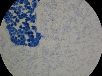


The Virtual Edge


Biol 230 Lab Manual Lab 1


Intestinal Colonization Of Genotoxic Escherichia Coli Strains Encoding Colibactin And Cytotoxic Necrotizing Factor In Small Mammal Pets Sciencedirect


Protocol For Hela Cells Infection With Escherichia Coli


Flower Like Patterns In Multi Species Bacterial Colonies Elife


Part a K Parts Igem Org


Observing Bacteria Under The Light Microscope Microbehunter Microscopy
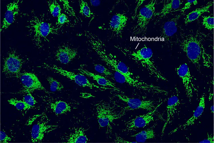


What Living Things You Can See Under A Light Microscope Rs Science


Flower Like Patterns In Multi Species Bacterial Colonies Elife
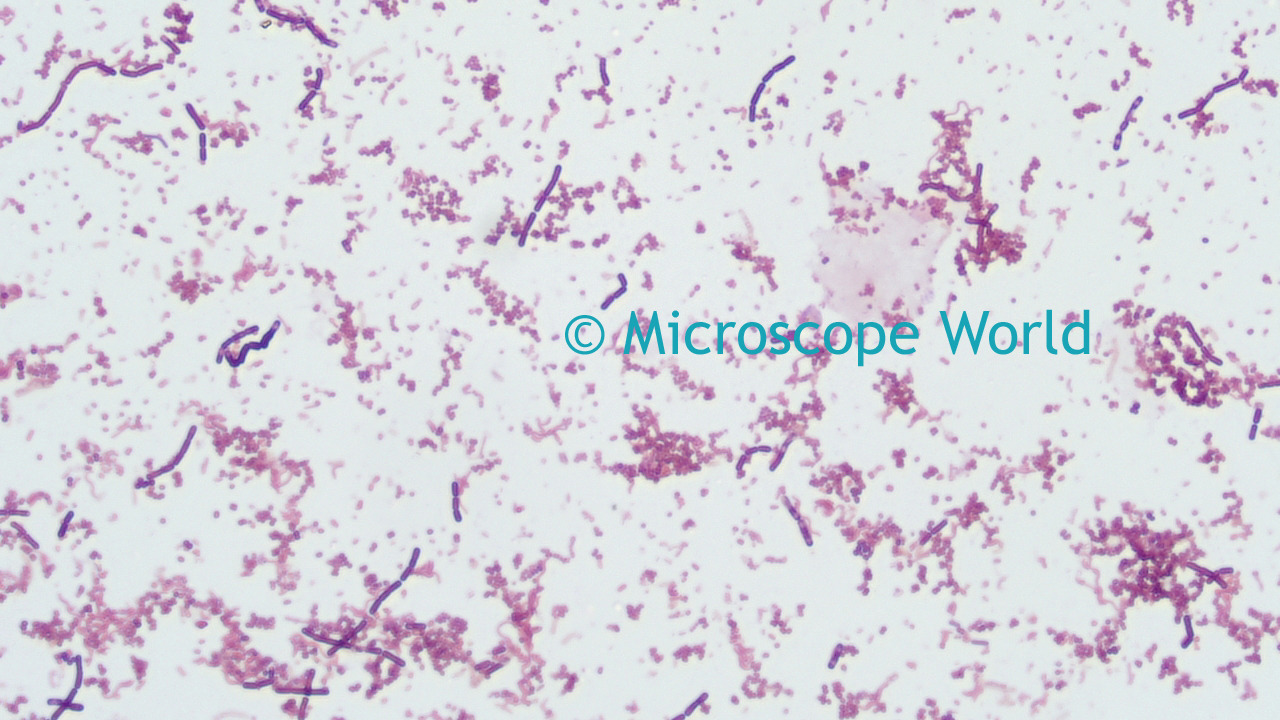


Microscope World Blog Microscopy Gram Staining


Lab Manual Exercise 1


Microscopic Studies Of Various Organisms


Spatial And Temporal Organization Of Reca In The Escherichia Coli Dna Damage Response Elife
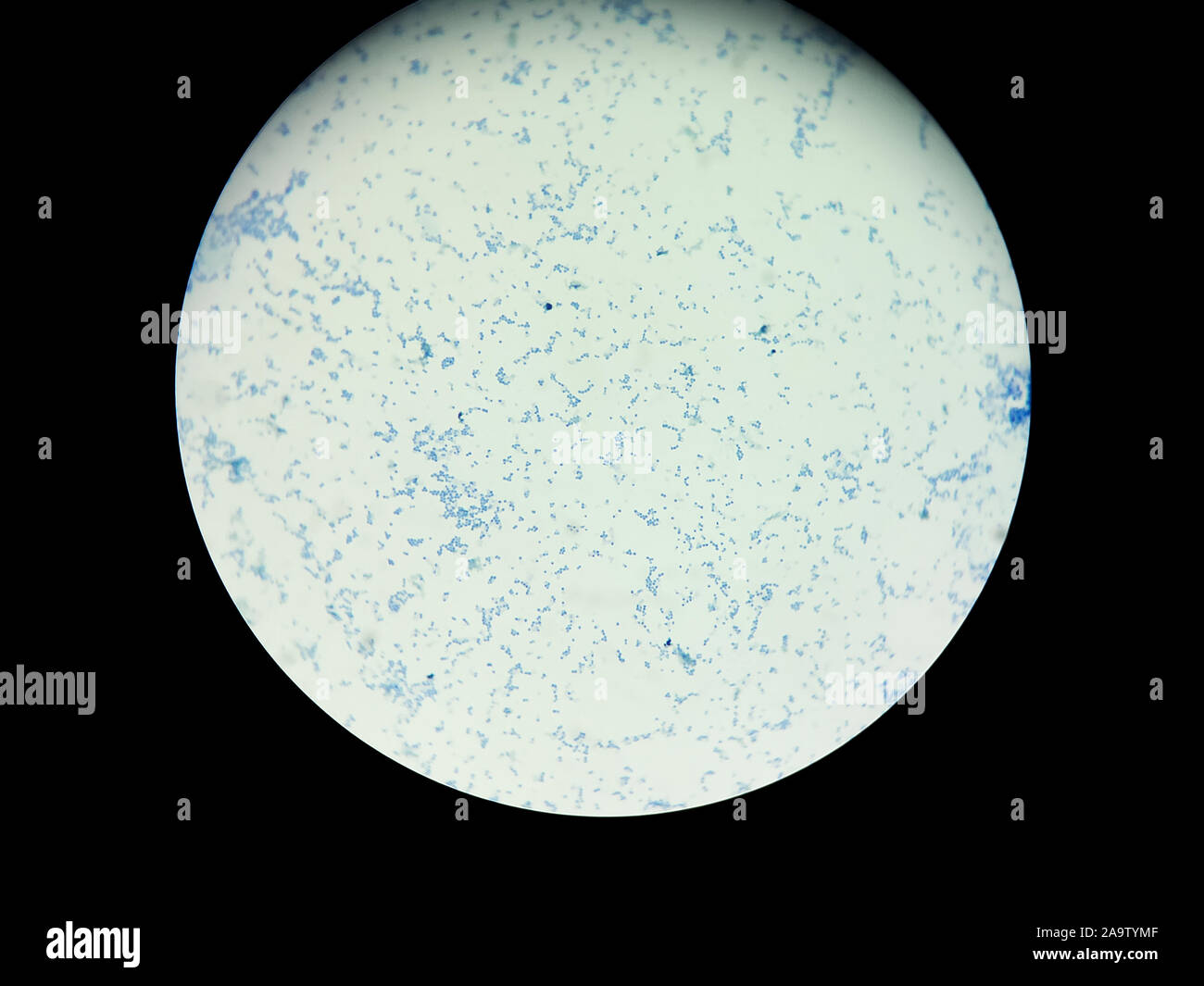


Light Microscope High Resolution Stock Photography And Images Alamy


Microscopy And Staining


What Does An E Coli Bacteria Look Like Under A Microscope Quora


Staphylococcus Aureus Under Microscope Microscopy Of Gram Positive Cocci Morphology And Microscopic Appearance Of Staphylococcus Aureus S Aureus Gram Stain And Colony Morphology On Agar Clinical Significance


Www Mccc Edu Hilkerd Documents Bio1lab3 Exp 4 000 Pdf


Microscope World Blog June 15


Gram Stain


Lab 1


Secreted Autotransporter Toxin Sat Induces Cell Damage During Enteroaggregative Escherichia Coli Infection


Gram Negative Synergy And Mechanism Of Action Of Alkynyl Bisbenzimidazoles Scientific Reports


Microbiology Lp1 Flashcards Quizlet


What Does An E Coli Bacteria Look Like Under A Microscope Quora


Microscope Observation Of Biofilm Inhibition Biofilm Inhibition Of E Download Scientific Diagram


What Magnification Do I Need To See Bacteria Westlab


Flower Like Patterns In Multi Species Bacterial Colonies Elife


E Coli Gram Stain Introduction Principle Procedure And Result Interpret


Please Answer From Penicilium To The End Thanks Fi Chegg Com


Biol 230 Lab Manual Lab 1


Can Nanoparticles Be Viewed Under Phase Contrast Microscope


Www Dechra Us Com Files Files Supportmaterialdownloads Us Us 077 Pra Pdf
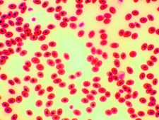


Microscope Images At Various Magnifications


Endospore Staining Principle Procedure And Results Learn Microbiology Online


Q Tbn And9gcs3nlev0tx1pfsddwm6y9ajujk4lxjzmg7ksdxctgnipv0l50c Usqp Cau


Microscopic Studies Of Various Organisms


Gram Stain


Escherichia Coli Bacteria E Coli Stock Footage Video 100 Royalty Free Shutterstock


Lab Manual Exercise 1


Gram Stain
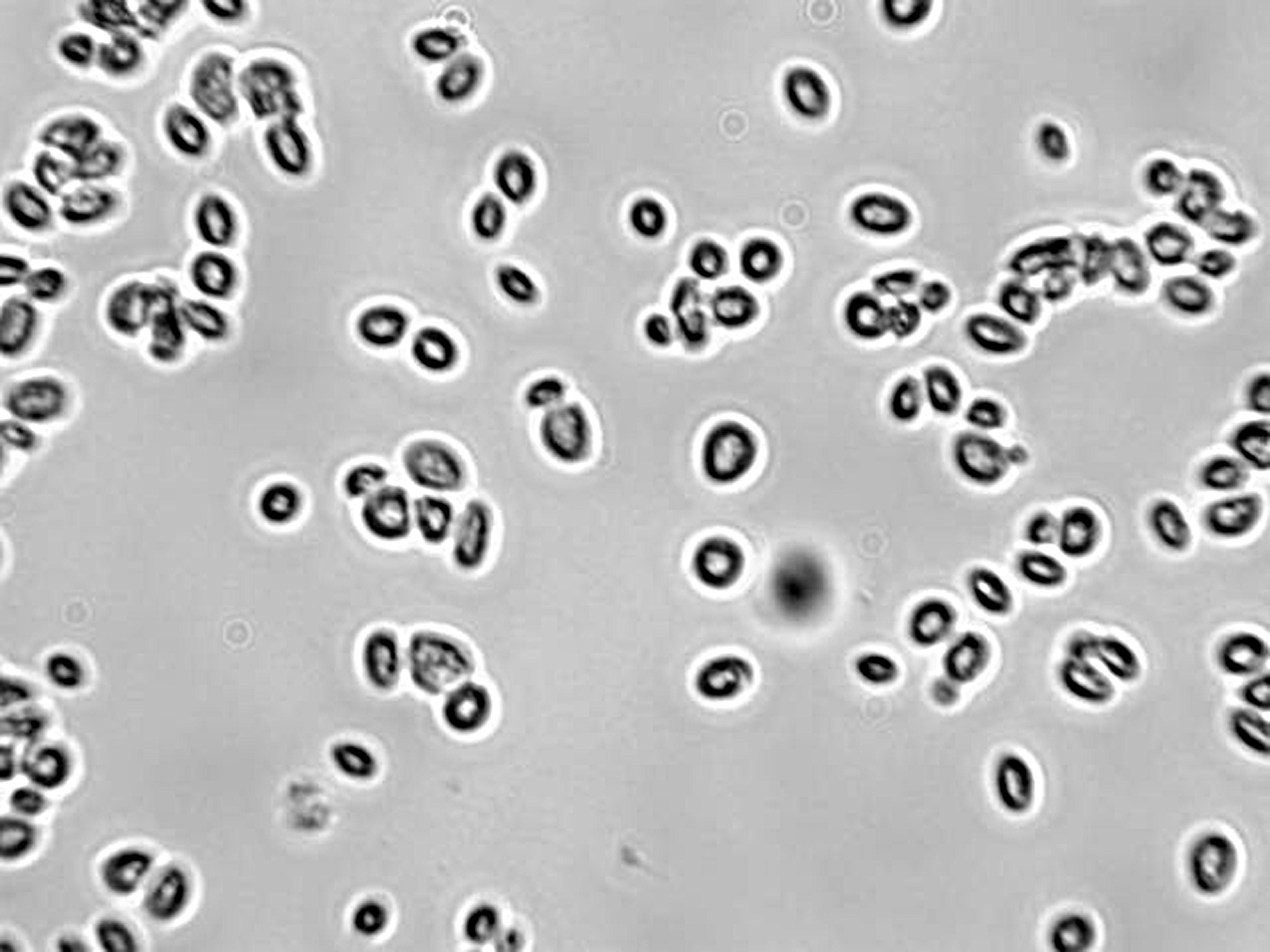


Microscopy For The Winery Viticulture And Enology
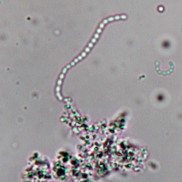


Observing Bacteria Under The Light Microscope Microbehunter Microscopy
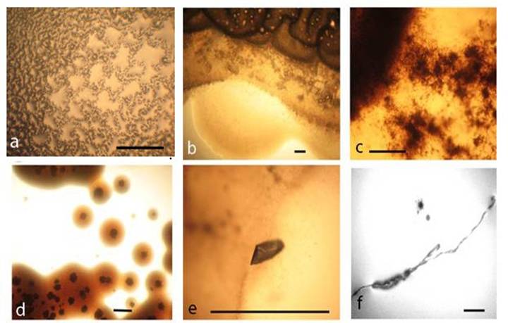


Survival Of Escherichia Coli Under Lethal Heat Stress By L Form Conversion


Biol 230 Lab Manual Lab 1


Bacteria Primed By Antimicrobial Peptides Develop Tolerance And Persist Biorxiv


Lab 1


Staphylococcus Aureus Under Microscope Page 1 Line 17qq Com
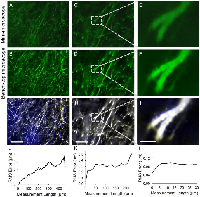


Hybrid Microscopy Enabling Inexpensive High Performance Imaging Through Combined Physical And Optical Magnifications Scientific Reports
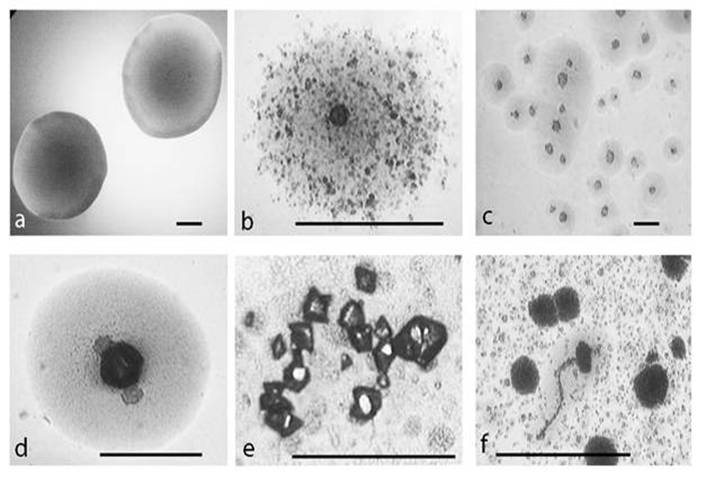


Survival Of Escherichia Coli Under Lethal Heat Stress By L Form Conversion


Team Leicester August12 12 Igem Org


What Does An E Coli Bacteria Look Like Under A Microscope Quora
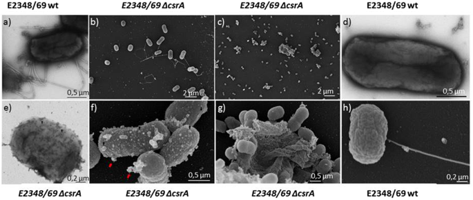


Metabolome And Transcriptome Wide Effects Of The Carbon Storage Regulator A In Enteropathogenic Escherichia Coli Scientific Reports


Microscopy Gram Staining Microscope World Blog
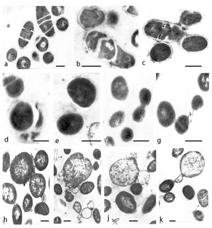


Survival Of Escherichia Coli Under Lethal Heat Stress By L Form Conversion


Observing Bacteria Under The Light Microscope Microbehunter Microscopy


Microscopic Studies Of Various Organisms


What Does An E Coli Bacteria Look Like Under A Microscope Quora


Persister Cells Resuscitate Using Membrane Sensors That Activate Chemotaxis Lower Camp Levels And Revive Ribosomes Biorxiv


コメント
コメントを投稿